
Robotic Cornual Resection for Heterotopic Cornual Pregnancy in Twin Gestation: Techniques, Risks and Outcomes

A heterotopic cornual pregnancy in the setting of a monochorionic diamniotic twin gestation is one of the most demanding scenarios in early pregnancy care. It combines an ectopic implantation at a highly vascular uterine site with the need to protect an ongoing intrauterine twin pregnancy. The clinical window is narrow, the haemorrhage risk is real, and management requires coordinated decision-making between reproductive medicine, maternal foetal medicine, anaesthesia, radiology and minimally invasive gynaecological surgery.
Cornual and interstitial pregnancies are often discussed together, but the terms are not always used consistently. In practice, the key concern is implantation in the uterine cornual region, close to the tubal ostium and myometrial blood supply. When this occurs alongside a viable intrauterine pregnancy, systemic medical treatment is usually unsuitable because the intrauterine pregnancy is intended to continue. Surgery then becomes a method of definitive treatment, haemorrhage prevention and uterine preservation.

Understanding heterotopic cornual pregnancy in twin gestation
Heterotopic pregnancy refers to the simultaneous presence of an intrauterine pregnancy and an ectopic pregnancy. It was once considered extremely rare in spontaneous conception, but assisted reproductive technologies have increased recognition of the condition. Risk factors include ovulation induction, embryo transfer, previous ectopic pregnancy, tubal surgery, pelvic inflammatory disease and multiple gestation.
A cornual or interstitial ectopic pregnancy is especially hazardous because it can enlarge before rupture. The surrounding myometrium allows more expansion than the ampullary tube, while the vascular supply can produce rapid intraperitoneal bleeding once rupture occurs.
In a monochorionic diamniotic twin gestation, the complexity rises further. These pregnancies already carry higher risks than singleton pregnancies, including miscarriage, preterm birth, twin-to-twin transfusion syndrome and growth discordance. Adding a cornual ectopic pregnancy introduces several urgent questions:
Can the ectopic pregnancy be removed without compromising the intrauterine twins?
Is the uterine wall strong enough after repair to support continuing gestation?
How can blood loss be limited while avoiding agents harmful to the intrauterine pregnancy?
What surveillance is needed after surgery?
Diagnosis usually depends on high-quality transvaginal ultrasonography. Features may include an empty uterine cavity in simple ectopic cases, but in heterotopic pregnancy the intrauterine pregnancy can distract from the ectopic component. Suspicion should remain high when pain, adnexal tenderness, free fluid or an eccentric cornual mass is present despite a confirmed intrauterine pregnancy.
MRI can help when ultrasound is inconclusive, especially in stable patients. Serial haemoglobin, blood group and crossmatch, baseline renal and liver function, and careful anaesthetic assessment are part of preparation. The threshold for intervention is low when there are signs of impending rupture.
Why robotic surgery has a role in this setting
Conventional laparoscopy remains a proven approach for many ectopic pregnancies. Robotic surgery may offer specific technical advantages in cornual resection, particularly when the aim is precise excision and meticulous uterine reconstruction.
The appeal of ROBOTIC CORNUAL RESECTION lies in three features: stable three-dimensional vision, articulated instruments and tremor filtration. These can help when working around the cornual region, where the surgeon must balance adequate removal of trophoblastic tissue with conservation of healthy myometrium.
Robotic assistance may be useful when:
The cornual mass is large but unruptured.
The ectopic pregnancy is close to major uterine vessels.
Multilayer uterine closure is expected to be difficult.
Fertility preservation and continuation of the intrauterine pregnancy are priorities.
The surgeon has advanced robotic suturing experience.
That said, robotic surgery is not automatically superior. It takes time to dock the system, requires trained staff and may not suit unstable patients. In haemodynamic instability or rupture with major bleeding, immediate laparoscopy or laparotomy may be safer depending on local expertise and resources.
Preoperative planning should be deliberate and multidisciplinary
The best outcomes are usually linked to early diagnosis and careful preparation. A typical preoperative plan includes:
Confirmation of anatomy
A senior sonologist or maternal foetal medicine specialist should confirm the intrauterine twin gestation, chorionicity, amnionicity, cardiac activity and location of the ectopic mass. The relationship of the cornual pregnancy to the uterine cavity, tubal ostium and serosa guides the incision plan.
Counselling and consent
Consent should cover bleeding, transfusion, conversion to laparotomy, miscarriage, loss of one or both foetuses, hysterectomy in a life-threatening emergency and later uterine rupture. Patients should also understand that future delivery is often planned by caesarean section after full-thickness cornual repair.
Blood readiness
Cornual surgery can bleed quickly. Crossmatched blood, large-bore intravenous access and a clear massive haemorrhage protocol are prudent.
Pregnancy-preserving choices
Systemic methotrexate is avoided when a viable intrauterine pregnancy is to be preserved. Local injection techniques have been reported for selected ectopic pregnancies, but cornual heterotopic pregnancy with twin gestation often requires definitive removal because of rupture risk and diagnostic certainty.
Anaesthetic considerations
The anaesthetic team should minimise maternal hypoxia, hypotension and excessive uterine irritability. Pneumoperitoneum pressure is usually kept as low as practical while preserving visualisation.
Key steps in robotic cornual resection
The exact method varies with anatomy, gestational age, surgeon preference and platform. The following sequence reflects common principles used in reported minimally invasive approaches.
Position the patient and access the abdomen safely
The patient is placed in dorsal lithotomy with left tilt when appropriate. Sequential compression devices are applied. After induction of anaesthesia, abdominal entry is performed away from the enlarged uterus. Many surgeons use an optical trocar or open entry technique.
Port placement must allow distance from the uterus and comfortable robotic arm movement. The camera port is usually placed above the umbilicus, with lateral robotic ports under direct vision. An assistant port allows suction, specimen handling, needle introduction and rapid help if bleeding occurs.
Survey the pelvis and confirm operative findings
Before docking or after initial access, the surgeon assesses haemoperitoneum, pelvic adhesions, the uterine cornua, tubes and ovaries. The cornual pregnancy may appear as a bulging vascular mass in the lateral uterine fundus. The intrauterine pregnancy is not directly visible from the abdomen, so ultrasound findings remain central.
If there is active bleeding, the plan may shift from controlled resection to emergency haemostasis.
Reduce vascularity before incision
Haemostasis is the major technical challenge. Options include bipolar coagulation around the planned incision line, temporary vascular control, diluted vasopressin injection into the myometrium or early suturing around the cornual base. Each has trade-offs.
Vasopressin can reduce bleeding but may cause cardiovascular effects if injected intravascularly or in high doses. In pregnancy, the dose and concentration should be conservative, and anaesthesia must be alerted before use.
Energy application should be precise. Excess thermal damage may weaken myometrium and affect later healing.
Incise the cornual serosa and develop the plane
A linear or elliptical incision is made over the most prominent part of the cornual swelling. The goal is to enter the gestational sac or dissect around it while preserving as much healthy myometrium as possible.
The robotic wristed instruments help maintain traction and counter-traction. Suction keeps the field clear. The surgeon identifies trophoblastic tissue and separates it from the myometrial bed. If the ipsilateral fallopian tube is diseased or directly involved, salpingectomy may be performed.
Excise the ectopic pregnancy completely
The ectopic gestational tissue is removed en bloc when feasible. Fragmented removal may be necessary if the tissue is friable, but complete evacuation is essential to reduce persistent trophoblast.
All tissue should be placed in an endoscopic retrieval bag. The operative field is inspected for residual products, bleeding points and breach into the uterine cavity. If the uterine cavity is entered, closure must be especially secure.
Reconstruct the uterine wall in layers
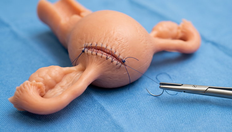
Uterine repair is the defining step. The surgeon closes the myometrial defect in two or more layers using delayed absorbable sutures. Barbed sutures may help maintain tension, although some surgeons prefer conventional sutures in pregnancy.
The closure should:
Obliterate dead space.
Approximate deep myometrium without strangulation.
Reconstruct the serosal surface.
Achieve haemostasis without excessive cautery.
Restore a contour that can tolerate uterine enlargement.
Robotic suturing can be valuable here because the cornual angle is awkward with straight-stick laparoscopy.
Confirm haemostasis and reassess the pregnancy
After irrigation and pressure reduction, the surgical bed is checked again. Even mild oozing can become significant later. Haemostatic agents may be used selectively.
Before leaving theatre or soon after recovery, ultrasound assessment confirms foetal cardiac activity in the ongoing twin pregnancy. Anti-D immunoglobulin is administered when indicated.
Potential benefits of the robotic approach
Robotic cornual surgery is attractive because it may combine the benefits of minimally invasive surgery with improved dexterity for reconstruction.
Potential benefits include:
Better suturing ergonomics
The cornual region is difficult to close securely. Robotic articulation can make deep myometrial suturing more controlled.
Reduced abdominal wall trauma
Compared with laparotomy, minimally invasive access may reduce postoperative pain, ileus and wound complications.
Clear visualisation
Three-dimensional magnified vision helps define tissue planes and bleeding points.
Faster recovery in selected patients
Early mobilisation and shorter hospital stay may be possible when blood loss is controlled and the intrauterine pregnancy remains stable.
Fertility-preserving repair
A careful multilayer closure supports uterine integrity for the continuing pregnancy and future reproductive planning.
These benefits depend heavily on case selection and surgical expertise. A robotic platform does not compensate for delayed diagnosis, poor haemorrhage planning or limited experience with cornual reconstruction.
Risks and limitations must be discussed clearly
The risks are substantial and should not be softened.
Maternal risks include haemorrhage, transfusion, conversion to laparotomy, injury to adjacent organs, infection, anaesthetic complications, thromboembolism and, rarely, hysterectomy. Persistent trophoblastic tissue can occur if excision is incomplete, requiring ongoing beta-hCG monitoring in cases where interpretation is possible despite intrauterine pregnancy.
Pregnancy-related risks include miscarriage, preterm labour, membrane rupture, foetal loss and complications related to anaesthesia or maternal instability. In monochorionic diamniotic twins, ongoing surveillance is already intensive, and surgery adds another layer of risk.
Later uterine rupture is a serious concern after full-thickness cornual surgery. Many teams recommend avoiding labour and planning caesarean delivery, often before the onset of contractions. The timing must be individualised with maternal foetal medicine input.
There are also system-level limitations. Robotic systems may not be available in all centres, including many parts of India. The cost can be high, emergency access may be limited, and outcomes depend on the entire theatre team.
What case reports and recent literature suggest
High-quality comparative trials are not available for this exact scenario because heterotopic cornual pregnancy with monochorionic diamniotic twins is exceptionally rare. Most evidence comes from case reports, small case series and extrapolation from laparoscopic management of interstitial pregnancy and robotic uterine surgery.
Reported cases of heterotopic interstitial or cornual pregnancy managed surgically show that continuation of the intrauterine pregnancy is possible after conservative excision and uterine repair. Many reports emphasise the same themes: early ultrasound diagnosis, avoidance of systemic methotrexate, rapid access to skilled minimally invasive surgery and careful postoperative obstetric surveillance.
Recent literature on robotic management of cornual and interstitial pregnancies describes successful resection with controlled blood loss in selected stable patients. The strongest technical advantage described is suturing. Robotic platforms allow precise multilayer closure, which is particularly relevant when preserving uterine strength matters.
Yet the evidence remains limited. Publications often involve single patients, varied gestational ages and different definitions of cornual versus interstitial pregnancy. Outcomes may be affected by publication bias, as successful rare cases are more likely to be reported. For clinicians and students, the correct interpretation is cautious optimism rather than broad generalisation.
A useful way to frame the evidence is this: robotic resection is a feasible option in expert hands for selected stable patients, but it is not the default answer for every cornual heterotopic pregnancy.
Postoperative care and follow-up
Postoperative management focuses on maternal stability, pregnancy viability and uterine healing.
Key steps include:
Serial vital signs and haemoglobin assessment.
Pain control compatible with pregnancy.
Early scan confirmation of twin cardiac activity.
Monitoring for contractions, bleeding or signs of infection.
Maternal foetal medicine follow-up for monochorionic surveillance.
Documentation of uterine incision site and repair technique.
Counselling about activity, warning symptoms and delivery planning.
Because monochorionic diamniotic twins need serial ultrasound surveillance, postoperative monitoring can be integrated with routine assessment for growth, amniotic fluid balance and Doppler findings. Cervical length assessment may also be considered depending on gestational age and symptoms.
The operative note should be detailed. Future obstetric teams need to know whether the uterine cavity was entered, how many suture layers were placed, what suture material was used and whether haemostatic agents or vascular control methods were applied.
Practical takeaway for medical teams
Robotic cornual resection for heterotopic cornual pregnancy in monochorionic diamniotic twin gestation sits at the intersection of emergency gynaecology, reproductive surgery and high-risk obstetrics. The procedure can be pregnancy-preserving, but only when diagnosis is timely, haemorrhage planning is rigorous and the surgical team can perform precise excision with secure uterine reconstruction.
For stable patients in centres with relevant expertise, robotic surgery may offer better dexterity and visualisation during one of the most technically demanding forms of ectopic pregnancy surgery. For unstable patients, speed and safety take priority over platform choice.
This article is for medical education only and does not replace specialist clinical judgement. Each case requires individualised assessment, informed consent and management by an experienced multidisciplinary team.


Comments