
Leiomyoma Care Integrating Phenotype Symptoms and Reproductive Goals for Precision Treatment

Leiomyomas are common, but their effects are rarely simple. One patient may have a small submucosal fibroid and severe bleeding. Another may have a large intramural fibroid and only mild pressure symptoms. A third may have no symptoms until infertility work-up reveals distortion of the uterine cavity.
This variation is why modern leiomyoma care is moving away from a single diagnosis and towards a more precise clinical model. The best treatment plan connects four factors: fibroid phenotype, symptom pattern, reproductive goals, and treatment risk.
Leiomyomas, also called uterine fibroids, are benign smooth muscle tumours of the uterus. They are among the most common gynaecological conditions in reproductive-age women. By late reproductive age, a large proportion of women will have at least one fibroid, though many remain asymptomatic. The clinical burden is still substantial. Fibroids contribute to heavy menstrual bleeding, anaemia, pelvic pain, urinary symptoms, infertility, pregnancy loss, caesarean birth, and reduced quality of life.
This article is for information only and does not replace individual medical advice, diagnosis, or treatment planning.
Leiomyomas are common, but the clinical burden is uneven
The prevalence of leiomyomas rises with age through the reproductive years. Many studies estimate that most women will develop fibroids by the time they reach menopause, although prevalence varies by ethnicity, diagnostic method, and access to imaging. Ultrasound-based screening detects many fibroids that never cause symptoms, while hospital-based data often over-represent patients with severe bleeding, pain, or fertility concerns.
The health impact is not limited to the uterus. Heavy menstrual bleeding can cause iron deficiency and anaemia, which may lead to fatigue, reduced exercise tolerance, poor concentration, and repeated healthcare visits. Bulk symptoms can affect bladder function, bowel habits, sexual health, and daily comfort. Pain can be cyclical or chronic. For some patients, the most distressing effect is reproductive uncertainty.
Fibroids also create system-level costs. They contribute to outpatient consultations, imaging, medical therapy, emergency visits for acute bleeding, blood transfusion, surgery, and time away from work or caregiving. In India and other settings where anaemia is already common, fibroid-related bleeding can intensify a major public health problem.
Yet the presence of a fibroid alone does not prove causation. Precise care begins by asking whether the fibroid’s site, size, number, and biology plausibly explain the symptoms.
Phenotype gives structure to a complex condition
A fibroid phenotype is the observable pattern of disease. It includes location, size, number, growth pattern, imaging appearance, molecular features where available, and clinical behaviour. Phenotyping helps clinicians move from “the patient has fibroids” to “this specific fibroid pattern is likely driving this specific problem”.
Location remains the most practical phenotype
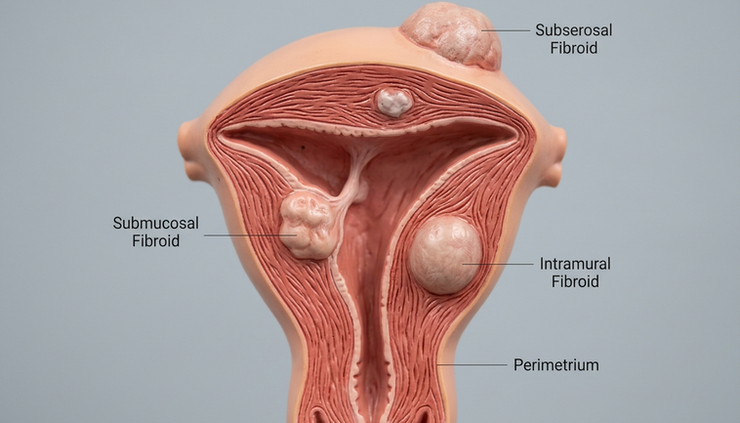
The International Federation of Gynecology and Obstetrics classification is widely used to describe fibroid relationship to the endometrium and serosa. In everyday practice, the most useful clinical categories are:
Phenotype | Common clinical pattern | Treatment implications |
Submucosal fibroids | Heavy menstrual bleeding, infertility, recurrent pregnancy loss when cavity distortion is present | Hysteroscopic myomectomy is often considered when feasible |
Intramural fibroids | Bleeding, pain, pressure, fertility impact if large or cavity-distorting | Options include medical therapy, myomectomy, ablation, embolisation, or hysterectomy depending on goals |
Subserosal fibroids | Bulk symptoms, pelvic pressure, urinary frequency | Fertility impact is usually lower unless anatomy is significantly altered |
Pedunculated fibroids | Pain from torsion may occur, mass effect possible | Surgical planning depends on stalk, size, and symptoms |
Multiple fibroids | Mixed symptoms and higher recurrence risk after uterus-sparing treatment | Requires careful mapping and counselling |
This classification does not answer every question, but it improves decision-making. A 2 cm submucosal fibroid may matter more for bleeding and implantation than a 5 cm subserosal fibroid. A uterus enlarged by multiple intramural fibroids may require a different plan from a single accessible lesion.
Biology is becoming part of the phenotype
Recent research has strengthened the view that leiomyomas are not one disease at the molecular level. Common genetic and molecular subtypes include fibroids with MED12 mutations, HMGA2 overexpression, and changes related to fumarate hydratase deficiency. These subtypes differ in extracellular matrix production, growth behaviour, and sometimes imaging appearance.
At present, molecular testing is not routine for most patients with typical fibroids. Still, this research matters. It explains why fibroids behave differently across patients and why future therapies may target specific pathways rather than only suppress ovarian hormones or remove tissue.
Researchers are also studying the fibroid microenvironment, progesterone signalling, angiogenesis, inflammation, vitamin D pathways, and extracellular matrix deposition. These areas may guide future treatment, especially for patients with repeated recurrence or poor response to standard therapies.
Symptoms should be mapped, not assumed
Fibroid care improves when clinicians separate symptoms into categories and match them to anatomy.
Heavy menstrual bleeding is the most common symptom that brings patients to care. Submucosal fibroids and cavity-distorting intramural fibroids are frequent contributors, but adenomyosis, ovulatory dysfunction, endometrial pathology, bleeding disorders, thyroid disease, and anticoagulant use may also play a role. A complete evaluation prevents overtreatment of incidental fibroids.
Bulk symptoms need different questioning. Urinary frequency, nocturia, constipation, bloating, pelvic heaviness, and lower back discomfort may reflect uterine size or fibroid position. Imaging should assess not only the largest diameter, but also the relationship to bladder, rectum, cervix, and pelvic sidewall.
Pain requires careful assessment. Fibroids can cause dysmenorrhoea, degeneration pain, or pressure-related discomfort. Acute severe pain may occur with torsion of a pedunculated fibroid or degeneration, especially in pregnancy. Chronic pain may also involve endometriosis, adenomyosis, pelvic floor dysfunction, or gastrointestinal causes.
Fertility and pregnancy-related symptoms need a separate frame. Cavity distortion has the clearest link with reduced fertility and miscarriage risk. The effect of non-cavity-distorting intramural fibroids is less clear and may depend on size, number, and associated uterine changes. Subserosal fibroids alone generally show less impact on fertility outcomes.
The key clinical question is not “Are fibroids present?” It is “Which fibroid is likely causing which symptom, and what will treatment change?”
Reproductive goals should guide the treatment pathway
Treatment planning must begin with the patient’s reproductive timeline. A 29-year-old planning pregnancy within a year needs different counselling from a 44-year-old who has completed childbearing and wants definitive relief from bleeding. Both deserve effective treatment, but the risk-benefit balance differs.
When pregnancy is desired soon
For patients trying to conceive or planning assisted reproduction, cavity evaluation is central. Saline infusion sonography, hysteroscopy, or high-quality transvaginal ultrasound can clarify whether a fibroid distorts the endometrial cavity. MRI may help when fibroids are multiple, large, or difficult to map.
Hysteroscopic myomectomy is often preferred for accessible submucosal fibroids. For selected intramural fibroids, abdominal, laparoscopic, or robotic myomectomy may be considered, especially when there is cavity distortion, repeated pregnancy loss, or failed fertility treatment with no other explanation.
Medical therapies that suppress ovulation or shrink fibroids temporarily may improve anaemia before surgery, but they do not usually serve as stand-alone fertility treatments. GnRH agonists or antagonists can reduce bleeding and volume for a defined period, though symptom recurrence after stopping is common.
When pregnancy is possible but not immediate
Some patients want uterine preservation but are not trying to conceive soon. In this group, the plan should balance symptom control, recurrence risk, and future options. Medical therapy may be appropriate for bleeding control, especially when symptoms are moderate and fibroids do not severely distort the uterus.
Options may include:
Tranexamic acid during menses for heavy bleeding
Non-steroidal anti-inflammatory medicines for pain in suitable patients
Hormonal contraceptives for cycle control
Levonorgestrel intrauterine system when the cavity shape allows safe placement
GnRH antagonist therapy with add-back treatment where available and appropriate
Short-term GnRH agonist therapy, often before surgery or to correct anaemia
Clinicians should document whether a treatment preserves fertility, delays definitive care, or may complicate future uterine surgery.
When childbearing is complete
For patients who do not desire future pregnancy, the full range of options can be discussed. Uterine artery embolisation may provide symptom relief for selected patients, especially with bulk and bleeding symptoms, though counselling should cover post-procedure pain, possible reintervention, and fertility uncertainty.
Radiofrequency ablation and focused ultrasound are uterus-sparing options in selected cases, depending on lesion size, number, and location. Availability varies by region and facility.
Hysterectomy remains the definitive treatment for symptomatic fibroids when fertility is not desired and less invasive options are unsuitable, ineffective, or unwanted. It eliminates recurrence but requires careful counselling about surgical route, ovarian conservation, recovery, and patient preferences.

Recent research is changing the treatment conversation
Several areas of recent research are shaping clinical practice.
Newer oral GnRH antagonists with hormonal add-back therapy have expanded non-surgical treatment for moderate to severe fibroid symptoms. Studies show that these medicines can reduce heavy menstrual bleeding and improve anaemia in many patients while limiting hypoestrogenic adverse effects through add-back therapy. They are not a permanent cure, but they can be useful for symptom control, preoperative preparation, or bridging to menopause in selected patients.
Imaging research has also improved treatment selection. MRI can identify fibroid number, location, vascularity, degeneration, and features that may influence response to embolisation or ablation. It also helps distinguish adenomyosis, which often coexists with fibroids and can explain persistent bleeding or pain after fibroid-targeted treatment.
Genomic research continues to refine phenotype categories. MED12-mutated fibroids appear common, while HMGA2-related fibroids may show different growth and tissue characteristics. Fumarate hydratase-related fibroids are less common but clinically relevant because they may be associated with hereditary syndromes in selected patients, especially when fibroids are numerous, early-onset, or have unusual pathology. Providers should follow local referral pathways when hereditary risk is suspected.
Research on racial and ethnic disparities has also shown that fibroid burden, age of onset, symptom severity, and access to minimally invasive care differ across populations. Precision treatment must include equitable diagnosis, counselling, and referral, not only advanced technology.
Practical recommendations for healthcare providers
Precision begins with a consistent clinical workflow. The following steps can improve counselling and reduce mismatched treatment.
Build a structured symptom profile
Ask about bleeding volume, cycle regularity, clots, flooding, pain timing, pressure symptoms, urinary and bowel symptoms, sexual pain, fatigue, and anaemia symptoms. Use haemoglobin and ferritin testing when heavy bleeding is present, especially in settings where iron deficiency is common.
Map the uterus before choosing treatment
Document fibroid number, maximum size, FIGO type or location, cavity distortion, uterine volume, and coexisting adenomyosis if suspected. Transvaginal ultrasound is often sufficient as first-line imaging. MRI is useful when anatomy is complex or when planning embolisation, ablation, or extensive surgery.
Align treatment with reproductive goals
Record whether the patient desires future pregnancy, is actively trying to conceive, is using assisted reproduction, wants uterine preservation without pregnancy, or has completed childbearing. This one step can prevent inappropriate use of procedures that may compromise future fertility.
Treat anaemia early
Do not wait until surgery to correct iron deficiency. Oral or intravenous iron may be needed depending on severity, tolerance, timeline, and local protocols. Correcting anaemia improves safety and quality of life.
Discuss recurrence and reintervention
Uterus-sparing treatment can be effective, but new fibroids or regrowth can occur. Patients should understand the chance of needing repeat treatment, especially when fibroids are multiple or treatment occurs at a younger age.
Use shared decision-making with clear language
Explain likely benefits, recovery time, effect on fertility, need for contraception during medical therapy, pregnancy timing after surgery, and possible complications. Good counselling compares realistic options rather than presenting one default treatment.
Precision care means treating the right problem
Leiomyomas deserve a more refined approach than “watch, medicate, or operate”. The best care connects phenotype with the symptom that matters most, then filters treatment through reproductive goals and individual risk.
For healthcare providers, the practical shift is clear: classify the fibroid pattern, confirm symptom causality, ask about pregnancy goals early, correct anaemia, and choose therapy that fits the person’s timeline. That approach makes leiomyoma care more precise, more respectful, and more likely to deliver outcomes that patients can actually feel.


Comments