Integrating Vascular Insights into Cesarean Closure Strategies for Optimal Outcomes
- Dr. Reena Sherene
- Jun 28
- 3 min read

Cesarean delivery remains one of the most common surgical procedures worldwide. Despite advances in surgical techniques, complications related to uterine healing and blood supply continue to challenge obstetricians. Understanding the vascular anatomy and perfusion patterns of the uterus, especially the lower uterine segment, can significantly influence cesarean closure strategies and improve patient outcomes. This post explores the intricate blood supply dynamics, the three-dimensional architecture of uterine and ovarian arteries, and how these insights inform surgical decisions.
Blood Supply Dynamics of the Lower Uterine Segment
The lower uterine segment (LUS) undergoes significant changes during pregnancy, becoming the primary site for cesarean incisions. Its blood supply differs from the upper segment, which affects healing and oxygen delivery after surgery.
Primary arterial sources: The LUS receives blood mainly from branches of the uterine artery, which itself arises from the internal iliac artery. The ovarian artery also contributes via anastomoses.
Collateral circulation: Rich collateral networks exist between uterine and ovarian arteries, ensuring continuous perfusion even if one source is compromised.
Vascular density: The LUS has a lower vascular density compared to the upper segment, which may influence tissue oxygenation and repair after cesarean closure.
These dynamics highlight the importance of preserving vascular integrity during cesarean delivery to support optimal healing.
Three-Dimensional Architecture of Uterine and Ovarian Arterial Systems
The uterine and ovarian arteries form a complex three-dimensional network that supports the uterus throughout pregnancy and postpartum.
Uterine artery: This artery courses medially along the broad ligament, giving off arcuate arteries that penetrate the myometrium. These further branch into radial and spiral arteries supplying the endometrium.
Ovarian artery: Originating from the abdominal aorta, it travels through the suspensory ligament of the ovary and anastomoses with uterine artery branches near the uterine cornu.
3D vascular arrangement: The intertwining of these arteries creates a mesh-like network that adapts dynamically to the growing uterus, ensuring adequate blood flow even under mechanical stress.
Understanding this 3D architecture helps surgeons anticipate areas of high vascularity and avoid excessive bleeding during uterine incision and closure.
Perfusion Patterns and Their Implications for Cesarean Delivery
Perfusion patterns in the uterus vary between segments and throughout pregnancy, influencing surgical outcomes.
Upper uterine segment: Exhibits robust blood flow with high oxygen delivery, supporting rapid healing.
Lower uterine segment: Shows comparatively reduced perfusion, which can delay tissue repair and increase risk of scar defects.
Impact on closure technique: Surgeons must consider these differences when selecting suture methods and materials. For example, minimizing trauma to the LUS vasculature can preserve perfusion and reduce complications such as uterine rupture or dehiscence.
Monitoring perfusion intraoperatively, possibly with Doppler ultrasound, can guide real-time decisions to optimize closure.
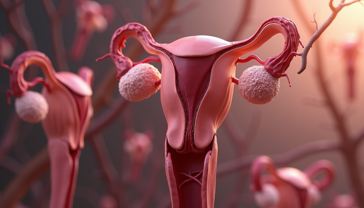
Significance of Vascular Casts in Understanding Uterine Blood Supply
Vascular casting is a technique where resin or other materials fill blood vessels, creating a detailed mold of the vascular network. This method has provided valuable insights into uterine blood supply.
Visualization of microvasculature: Vascular casts reveal the fine branching patterns and density of vessels in the LUS and upper segment.
Identification of anastomoses: Casts demonstrate the extent of connections between uterine and ovarian arteries, highlighting potential collateral pathways.
Surgical planning: By understanding these patterns, surgeons can better predict bleeding risks and plan incisions to avoid major vessels.
Research using vascular casts has advanced knowledge of uterine perfusion, directly impacting cesarean closure techniques.
Differences in Oxygen Delivery Post-Surgery Between Lower and Upper Uterine Segments
Oxygen delivery is critical for tissue repair after cesarean section. The LUS and upper segment differ significantly in this regard.
Lower uterine segment: Due to its lower vascular density and perfusion, oxygen delivery is reduced. This can slow healing and increase scar vulnerability.
Upper uterine segment: Maintains higher oxygen levels, supporting faster and more robust tissue regeneration.
Clinical implications: Surgeons should consider these differences when closing the uterus. Techniques that preserve blood flow and minimize ischemia in the LUS can reduce postoperative complications.
For example, avoiding excessive electrocautery and using interrupted sutures may help maintain better oxygenation in the LUS.
Reference:
AJOG expert review: Beyound Sutures: incorporating vascular insights into cesarean closure discussions-Blood supply for the LUS


Comments